Skip to content
  • Home
  • About Us
  • Our Services
    • Gynaecological Ultrasound
    • Breast Ultrasound
    • Echocardiography
    • Insurance Examinations
    • Musculoskeletal Ultrasound
    • Pregnancy Ultrasound
    • Doppler Ultrasound
    • Ultrasound
    • Workers Compensation
  • Ultrasound
    • Breast Ultrasound
    • Doppler Ultrasound
    • Obstetrics Ultrasound
    • Musculoskeletal Ultrasound
    • Echocardiogram Ultrasound
    • General Ultrasound
    • Workers Compensation
  • FAQs
  • For Referrers
    • NRI Referral
    • Referrer Registration
    • Imaging Portal
  • Contact Us
Menu
  • Home
  • About Us
  • Our Services
    • Gynaecological Ultrasound
    • Breast Ultrasound
    • Echocardiography
    • Insurance Examinations
    • Musculoskeletal Ultrasound
    • Pregnancy Ultrasound
    • Doppler Ultrasound
    • Ultrasound
    • Workers Compensation
  • Ultrasound
    • Breast Ultrasound
    • Doppler Ultrasound
    • Obstetrics Ultrasound
    • Musculoskeletal Ultrasound
    • Echocardiogram Ultrasound
    • General Ultrasound
    • Workers Compensation
  • FAQs
  • For Referrers
    • NRI Referral
    • Referrer Registration
    • Imaging Portal
  • Contact Us
Referrer's Login
Book an Appointment
Menu
  • Home
  • About Us
  • Our Services
    • Gynaecological Ultrasound
    • Breast Ultrasound
    • Echocardiography
    • Insurance Examinations
    • Musculoskeletal Ultrasound
    • Pregnancy Ultrasound
    • Doppler Ultrasound
    • Ultrasound
    • Workers Compensation
  • Ultrasound
    • Breast Ultrasound
    • Doppler Ultrasound
    • Obstetrics Ultrasound
    • Musculoskeletal Ultrasound
    • Echocardiogram Ultrasound
    • General Ultrasound
    • Workers Compensation
  • FAQs
  • For Referrers
    • NRI Referral
    • Referrer Registration
    • Imaging Portal
  • Contact Us

Echocardiography

Home / Services / Echocardiography

Contents

Other Services

More Services
doctor-performing-ultrasound-scanning-her-pregnant-patient

Gynaecological Ultrasound

gynecologist-performing-ultrasound-consultation

Doppler Ultrasound

young-woman-getting-ultrasound-scanning-examination-hospi

Breast Ultrasound

gynecologist-performing-ultrasound-consultation

Ultrasound

health-benefits-claim-benefits-form-concept

Insurance Examinations

physical-therapist-using-ultrasound-probe-patient-s-shoulder-hospital

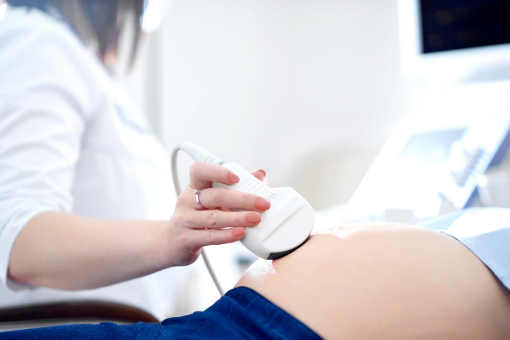
An echocardiogram, often referred to in the medical community as a cardiac ECHO or simply an ECHO, is a ultrasound of the heart. Also known as a cardiac ultrasound, it uses standard ultrasound techniques to image two-dimensional slices of the heart. The latest ultrasound systems that ARC XRay & Imaging now employ utilises 3D real-time imaging.

In addition to creating two-dimensional pictures of the cardiovascular system, an echocardiogram can also produce accurate assessment of the velocity of blood and cardiac tissue at any arbitrary point using pulsed or continuous wave Doppler ultrasound. This allows assessment of cardiac valve areas and function, any abnormal communications between the left and right side of the heart, any leaking of blood through the valves (valvular regurgitation), and calculation of the cardiac output as well as the ejection fraction.

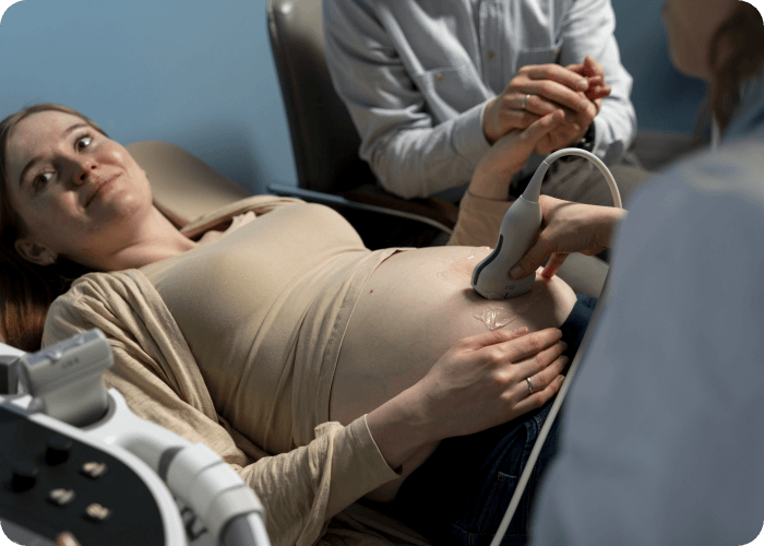
Echocardiography is performed by cardiac sonographers.

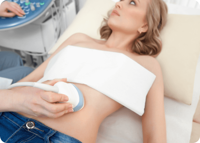
Preparation: You will be required put on a robe. There is no other preparation required. The examination takes approximately 30-45 minutes.

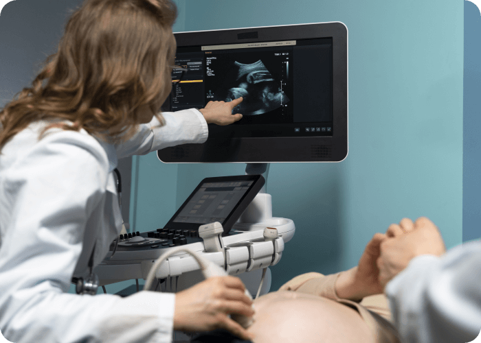
Echocardiography is used to diagnose cardiovascular diseases. In fact, it is one of the most widely used diagnostic tests for heart disease. It can provide a wealth of helpful information, including the size and shape of the heart, its pumping capacity and the location and extent of any damage to its tissues. It is especially useful for assessing diseases of the heart valves. It not only allows doctors to evaluate the heart valves, but it can detect abnormalities in the pattern of blood flow, such as the backward flow of blood through partly closed heart valves, known as regurgitation. By assessing the motion of the heart wall, echocardiography can help detect the presence and assess the severity of coronary artery disease, as well as help determine whether any chest pain is related to heart disease. Echocardiography can also help detect hypertrophic cardiomyopathy. The biggest advantage to echocardiography is that it is noninvasive (doesn’t involve breaking the skin or entering body cavities) and has no known risks or side effects.

Transthoracic echocardiogram

Main article: Transthoracic echocardiogram

A standard echocardiogram is also known as a transthoracic echocardiogram (TTE), or cardiac ultrasound. In this case, the echocardiography transducer (or probe) is placed on the chest wall (or thorax) of the subject, and images are taken through the chest wall. This is a non-invasive, highly accurate and quick assessment of the overall health of the heart.

Transoesophageal echocardiogram

This is an alternative way to perform an echocardiogram. A specialized probe containing an ultrasound transducer at its tip is passed into the patient’s oesophagus. This allows image and Doppler evaluation which can be recorded. This is known as a transoesophageal echocardiogram, or TOE (TEE in the United States).

3-dimensional echocardiography

3D echocardiogram of a heart viewed from the apex

3-D echocardiography is now possible, using an ultrasound probe with an array of transducers and an appropriate processing system. This enables detailed anatomical assessment of cardiac pathology, particularly valvular defects,[1] and cardiomyopathies.[2] The ability to slice the virtual heart in infinite planes in an anatomically appropriate manner and to reconstruct 3-dimensional images of anatomic structures make 3D echocardiography unique for the understanding of the congenitally malformed heart.

Other Services

doctor-performing-ultrasound-scanning-her-pregnant-patient

Gynaecological Ultrasound

gynecologist-performing-ultrasound-consultation

Doppler Ultrasound

young-woman-getting-ultrasound-scanning-examination-hospi

Breast Ultrasound

gynecologist-performing-ultrasound-consultation

Ultrasound

health-benefits-claim-benefits-form-concept

Insurance Examinations

More Services

Stay Patient, We're Prepping Our Website.

As we prepare to launch our website, we invite you to join us on this exciting journey. The future of ultrasound is just around the corner, and we can't wait to share it with you.

Contact Us On (02) 66842354
footer-logo

Northern Rivers Ultrasound and Imaging is part of the Advanced Radiology Imaging Group.

We are located at 56 Stuart St Mullumbimby directly next door to Mullumbimby Medical Centre. We specialise in all forms of ultrasound including Echocardiography. Pensioner and Health Care Card holder examinations available. The clinic is a standalone practice with off street parking that can be accessed from the back of the property through McGougans Lane.

Quick Links

  • Home
  • About Us
  • Our Services
  • Ultra Sound
  • FAQs
  • Contact Us

Services

  • Doppler Ultrasound
  • Breast Ultrasound
  • Ultrasound
  • Insurance Examinations
  • More Services

Company

  • Email
  • admin@arcxray.com.au
  • admin@nrimaging.com.au
  • Mullumbimby
  • (02) 668 423 54
  • 56 Stuart St, Mullumbimby NSW 2482, Australia
  • Yamba
  • (02) 66 46 9338
  • 3-4/12 Clarence Street Yamba NSW 2464
  • FAX
  • (02) 4210 8848

COPYRIGHT 2025. Northern Rivers Ultrasound and Imaging. ALL RIGHTS RESERVED

  • Privacy Policy
  • Terms of Use