Ultrasound imaging, also called ultrasound scanning or sonography, involves exposing part of the body to high-frequency sound waves to produce pictures of the inside of the body. Ultrasound exams do not use ionizing radiation (as used in x-rays). Because ultrasound images are captured in real-time, they can show the structure and movement of the body's internal organs, as well as blood flowing through blood vessels.
Ultrasound imaging is a noninvasive medical test that helps physicians diagnose and treat medical conditions.
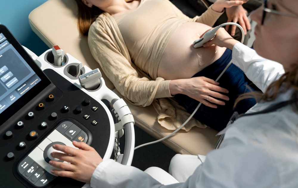
Obstetrical ultrasound provides pictures of an embryo or fetus within a woman's uterus.
A Doppler ultrasound study may be part of an obstetrical ultrasound examination.
Doppler ultrasound is a special ultrasound technique that evaluates blood velocity as it flows through a blood vessel, including the body's major arteries and veins in the abdomen, arms, legs and neck.
During an obstetrical ultrasound the examiner may evaluate blood flow in the umbilical cord or may in some cases assess blood flow in the fetus or placenta.